Purpose: The aim of this study was to identify the incidence of accessory roots in third molar teeth. Methods: This study evaluates the incidence of accessory roots present on maxillary and mandibular third molars in 4,904 teeth from a total pool of 1,492 patients, both male and female, ranging in age from fifteen to ninety-six years. The number of teeth containing an accessory root was recorded and correlations have been made comparing right to left third molars as well as maxillary to mandibular. The results were analyzed using a chi-squared test. Results: A total of 316 third molars were identified as having one or more accessory roots, representing an incidence of 6.43%. Of the 1,492 patients undergoing extraction of one or more third molar teeth, 239 (16%) of these patients had a tooth extracted that demonstrated evidence of accessory root formation. Conclusion: The high incidence of third molar teeth with one or more accessory roots provides evidence that may suggest an explanation for unexpected difficulty during a third molar extraction, and draws attention to the importance of thorough clinical and radiographic evaluation preoperatively, as well as to the need for effective risk management in third molar evaluation and treatment.
This is an Open Access article, distributed under the terms of the Creative Commons Attribution 4.0 International License (http://creativecommons.org/licenses/by/4.0/), which permits unrestricted use, distribution and reproduction in any medium or format, provided the original work is properly cited.
Throughout the evolutionary timeline of human development the anatomy of teeth has remained relatively constant especially when considering crown and root morphology
[1]
Nelson, S. J. Wheeler's dental anatomy, physiology and occlusion. (Elsevier Health Sciences, 2014).
[1]
. Given this, third molars in particular are a common exception to this consistency, often demonstrating significant variation in size, shape and presence even for a single patient
[2]
Carter, K. W., S. Morphologic and Demographic Predictors of Third Molar Agenesis A Systematic Review and Meta-analysis. Journal of dental research 94, 886-894 (2015).
[2]
. Accordingly, when performing diagnostic and surgical procedures on any third molar tooth, the practitioner can expect an increased degree of variability as to both crown and root morphology, as well as with the numbers of roots, though these variabilities are may not be suggested radiographically
[3]
Guerisoli, D. External and Internal Anatomy of Third Molars Braz Dent J 9, 91-94 (1998).
[3]
.
The objective of this study was to identify the presence of third molars with accessory root formation in order to determine the relative regularity of incidence, as well as to correlate accessory root development with anatomical location. Additionally, consideration was given to patients undergoing extraction of more than one third molar tooth in order to draw conclusions about the incidence of accessory root incidence in individual patients.
The most common expected root structure of maxillary molars involves three roots, a mesial-buccal, distobuccal, and palatal, whereas mandibular molars most commonly demonstrate two roots, one mesial and one distal
[4]
Vertucci, F. Root canal anatomy of the human permanent teeth. Oral surgery, oral medicine, oral pathology 58, 589-599 (1984).
[4]
. Accessory roots form in similar fashion to normal roots, and occur as a result of the ingrowth of processes from the root sheath of Hertwig. As such, the development of three-rooted mandibular molars results either from a bifurcated mesial root or an accessory distolingual root, while four-rooted maxillary molars commonly have either a bifurcated mesiobuccal root or an accessory palatal root
[5]
Kocsis, G. S. M., Antonia. Accessory root formation on a lower medial incisor. Oral surgery, oral medicine, oral pathology 68, 644-645 (1989).
[5]
.
Scheid et al suggest that accessory roots typically form in teeth whose roots form after birth, and that these roots are most likely the result of trauma, metabolic dysfunction, or pressure. The authors argue that third molars are the multirooted teeth that that are most likely to exhibit accessory roots, and this is consistent with what Nelson et al described as the tendency of third molars to show more variation in development than any other teeth in the mouth
[6]
Scheid, R. C. Woelfel's Dental Anatomy. (Lippincott Williams and Wilkins, 2012).
[6]
.
The present study expanded upon previous publications by utilizing a larger patient population (1492 patients) from which to examine data and draw conclusions. The importance of this study cannot be underestimated with respect to its clinical applications, in particular regarding what clinicians may expect during the diagnostic, treatment planning and surgical phases related to the removal of third molar teeth. When the astute dental practitioner recognizes the incidence of accessory root formation that can be expected during the removal of third molar teeth, then that surgeon will be in a better position to deliver appropriate care that can serve to improve outcomes as well as to attenuate the risk management issues inherent in third molar diagnosis and treatment.
2. Materials and Methods
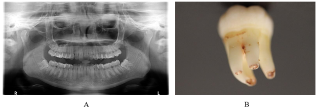
A total of 4904 third molar teeth were extracted from a total patient pool of 1492 individuals, ranging in age from 15 to 96 years old. Of the patients who were treated, 892 presented for extraction of all four third molar teeth. All patients were evaluated with a panoramic radiograph, either alone or in support of periapical radiographs, and all teeth were inspected following removal for the presence of one or more accessory roots. A patient was deemed acceptable for this study if root formation of the third molar was complete or nearly complete, which was judged to be approximately seventy-five per-cent of what one could reasonably expect to be complete root formation (Figure 1 A and B).
The results were then collated as to the percentage of third molar teeth either with or without one or more accessory roots, as well as with respect to the percentage of accessory root formation on each of the four numbered third molar teeth, i.e. #’s 1, 16, 17, and 32. Additionally, the percentage of patients with one or more accessory third molar root formation was tabulated. Finally, of the patients who underwent extraction of four third molar teeth, the percentage with one or more third molar tooth with accessory root formation was tabulated.
Figure 1. A. Panoramic Radiographs taken pre-operatively. Radiograph does not conclusively indicate the presence of an accessory root. B. The extracted mandibular left third molar tooth (#17) with two accessory roots present.
3. Results
Accessory roots were confirmed in 315 of the 4904 total number of third molar teeth examined in this study. This represents an incidence of 6.42%. Each third molar location was further analyzed to determine the site most likely to harbor a tooth with an accessory root. Of the third molars, tooth #32 demonstrated the highest likelihood of having an additional root. Of the 1,243 right mandibular third molars that were extracted 127 demonstrated accessory root formation, resulting in an incidence of 10.22%. The next most likely third molar to have accessory root formation was tooth #17, as of the 1,230 left mandibular third molars that were extracted 106 demonstrated accessory root formation, resulting in an incidence 8.62%. Maxillary molars exhibited significantly less accessory root formation, with tooth #1 demonstrating a 3.39% incidence and tooth #16 displaying a 3.36% incidence of accessory root formation. (Table 1)
Table 1. Incidence of accessory root formation in third molars by tooth number.
Tooth Number
Number of Teeth Extracted
Number of Accessory Roots
Incidence
1
1269
43
3.39%
16
1162
39
3.36%
17
1230
106
8.62%
32
1243
127
10.22%
Total
4904
315
6.42%
Accessory root formation within individual patients was also examined. It was determined that of the 1,492 individuals that underwent third molar extractions 239 of these patients demonstrated at least one third molar with an accessory root, representing an incidence of 16%. (Table 2)
Table 2. Incidence of accessory root formation in individual patients.
All Patients Presenting for At Least one 3rd molar extraction
Accessory Roots Present In At Least One Molar Tooth Extracted
Incidence
1492
239
16%
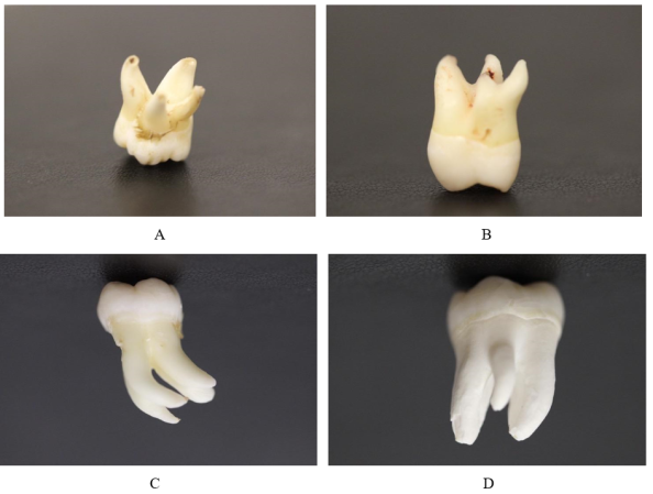
As noted above, there is variability as to the nature, location, and morphology of third molar accessory roots. Figure 2 demonstrates examples of maxillary third molar teeth with an accessory palatal root, as well a bifurcated mesiobuccal root. The mandibular teeth provide examples of bifurcated roots, as well as a smaller accessory root (Figure 2).
Table 3. Incidence among patients presenting for extraction of all four third molars.
Number of Patients Presenting for All Four 3rd Molars to be Extracted
Figure 2. Accessory root formation on extracted maxillary and mandibular third molars. A+B. Examples of accessory root formation on maxillary third molars. Complete and separate root accessory root formation shown in A. Root bifurcation in the apical third shown in B. B+C Examples of accessory root formation on mandibular third molars. Two complete accessory roots present in mandibular third molar shown in C. In D a separate but smaller accessory root is present.
Table 4. Combinations of third molars extracted with incidence of accessory roots.
Number of Patients with All Four 3rd Molars Extracted and >1 Tooth with an Accessory Root
Accessory Root on Both 17 & 32
Accessory Root on Both 1 & 16
Accessory Root on ¾ Teeth Extracted
Accessory Root on All 4 Teeth Extracted
58
34 (58.6%)
9 (15.5%)
5 (8.6%)
1 (1.7%)
As expected, patients who presented for removal of all four third molar teeth had an increased incidence of accessory roots. Of the 892 patients presenting for removal of four third molar teeth, 183 of these presented with an accessory root on one or more teeth, representing an incidence of 20.5%. Within this subset of patients, 125 patients (14%) demonstrated one third molar with an accessory root and 58 patients (6.5%) presented with more than one third molar with accessory roots (Table 3). Of these 58 patients, 34 (58.6%) demonstrated accessory roots on both #17 and #32, while nine patients (15.5%) demonstrated accessory roots on teeth #1 and #16. Of the remaining patients, five (8.6%) demonstrated accessory roots in three out of four third molars, while only one patient (1.7%) demonstrated accessory root formation in all four third molar teeth (Table 4).
4. Discussion
Several studies have been published highlighting the incidence of molars with an accessory root, but few have focused on accessory root formation among a large population that includes both maxillary and mandibular third molars specifically. The majority of these studies have focused on first molars, with very few studies solely dedicated to third molar morphology.
Ahmed et al published a strong review of accessory roots in maxillary molars in 2012. The article summarizes many previous reports of accessory roots, and categorizes the results based on individual tooth. The majority of the studies mentioned were case reports, though several observational studies were cited for maxillary third molars
[7]
Ahmed, H. M. & Abbott, P. V. Accessory roots in maxillary molar teeth: a review and endodontic considerations. Australian dental journal 57, 123-131; quiz 248,
. Of these studies, the report with the greatest number of teeth was 269. This study, published by Guerisoli et al in 1998, found that of 155 maxillary third molars examined, 5 (3.2%) demonstrated accessory root formation. This study also found that of 114 mandibular third molars only 1 (0.9%) showed formation of an accessory root
[3]
Guerisoli, D. External and Internal Anatomy of Third Molars Braz Dent J 9, 91-94 (1998).
[3]
. Of the other studies highlighted the most recent, a 2008 report by Cosic et al, reported that of 56 extracted maxillary third molars 1.8% demonstrated the presence of an accessory root
[8]
Cosic, J. Root Canal Anatomy of Third Molars PEF/IADR International Dental Research Meeting London (2008).
[8]
. This article also examined 50 mandibular third molars, of which none (0%) had an accessory root. Sidow et al reported the highest incidence in accessory roots in third molars; 5% (8 of 150) in mandibular third molars and 7% (11 of 150) in maxillary third molars
[9]
Sidow, S. J., West, L. A., Liewehr, F. R. & Loushine, R. J. Root canal morphology of human maxillary and mandibular third molars. J Endod 26, 675-678,
Although the accessory root is typically considered a variation it is not considered pathological. Indeed, the additional root has been regarded as a normal racial and morphological variation
[10]
Song, J. S. et al. Incidence and relationship of an additional root in the mandibular first permanent molar and primary molars. Oral surgery, oral medicine, oral pathology, oral radiology, and endodontics 107, e56-60,
. Several studies have been conducted that provide evidence to suggest that accessory roots are more common in Taiwanese, Indian, Korean, and Mongolianpopulations. As an example, Tu et al focused on three rooted mandibular first molars in Taiwanese individuals, and found an incidence of 33.33% among the 123 patients screened with CBCT
[11]
Tu, M. G. et al. Prevalence of three-rooted mandibular first molars among Taiwanese individuals. J Endod 33, 1163-1166,
Srivathsa, S. H. Prevalence of three rooted deciduous mandibular molars in Indian children. International Journal of Dental Science and Research 2, 14-16,
Song, J. S., Choi, H. J., Jung, I. Y., Jung, H. S. & Kim, S. O. The prevalence and morphologic classification of distolingual roots in the mandibular molars in a Korean population. J Endod 36, 653-657,
Ferraz, J. A. B. J. E. P. Three-rooted mandibular molars in patients of Mongolian, Caucasian and Negro origin. Braz J. 3, 113-117 (1992).
[15]
Tu, M. G. et al. Detection of permanent three-rooted mandibular first molars by cone-beam computed tomography imaging in Taiwanese individuals. J Endod 35, 503-507,
It is important to note that many of the studies that include accessory root statistics have been published from an endodontic perspective, and provide valuable information regarding both root and root canal morphology
[9]
Sidow, S. J., West, L. A., Liewehr, F. R. & Loushine, R. J. Root canal morphology of human maxillary and mandibular third molars. J Endod 26, 675-678,
Versiani, M. A., Pecora, J. D. & de Sousa-Neto, M. D. Root and root canal morphology of four-rooted maxillary second molars: a micro-computed tomography study. J Endod 38, 977-982,
. However, the present study focuses upon the incidence of accessory roots from the perspective of oral and maxillofacial surgery, specifically with respect to the surgical removal of third molar teeth. In particular, this study is intended to bring attention to the manner in which accessory root formation may influence diagnosis, treatment planning, and difficulty of surgery.
As such, given the relative incidence of accessory root formation in third molar teeth, the astute clinician should appreciate the potential complications that may arise during surgical removal due to the presence of one or more accessory roots. As the results of this study suggest, the practitioner can expect to encounter an accessory root approximately 3.37 percent of the time with maxillary third molars, approximately 9.42 percent of time with mandibular third molars, with an average percentage of 6.43 percent for all third molars. Additionally, when four third molar teeth are removed in one patient seating, the practitioner can expect to encounter multiple third molar teeth with accessory root formation approximately in 6.68 percent of these cases.
These results highlight the importance of thorough preoperative clinical and radiologic evaluation. While the most common preoperative radiograph utilized for the evaluation and treatment of third molar teeth is the panoramic radiograph, this radiographic modality provides only a two-dimensional visualization of tooth location and morphology, and as such it is not completely reliable with respect to identifying the presence of accessory roots associated with third molar teeth
[17]
Bell, G. W. e. a. The accuracy of dental panoramic tomographs in determining the root morphology of mandibular third molar teeth before surgery. Oral Surgery, Oral Medicine, Oral Pathology, Oral Radiology, and Endodontology 95, 119-125 (2003).
[17]
. It was suggested by Tu et al that CBCT (Cone-Beam Computed Tomography) could serve as a useful tool to reveal the true nature of tooth root structures in three dimensions
[15]
Tu, M. G. et al. Detection of permanent three-rooted mandibular first molars by cone-beam computed tomography imaging in Taiwanese individuals. J Endod 35, 503-507,
. Additionally, Guerrero et al’s recent study praises the CBCT for its ability to confirm the number of roots and root morphology, especially when compared to the standard panoramic radiograph
[18]
Guerrero, M. E. e. a. Can preoperative imaging help to predict postoperative outcome after wisdom tooth removal? A randomized controlled trial using panoramic radiography versus cone-beam CT. Clinical oral investigations 18, 335-342 (2014).
[18]
. Nevertheless, while the statistical evidence from this study suggest that CBCT may be useful diagnostic tool with respect to the identification of accessory root(s) on third molar teeth, the routine use of CBCT for third molar evaluation and treatment is not practical, and would be cost prohibitive. Additionally, although the removal of third molar teeth is a common procedure performed in dental practice, with a complication rate that is relatively low, it is reasonable to assume that a tooth with an additional root may increase the difficulty of any given extraction
[19]
Bui, C. H. S., Edward B.; Dodson, Thomas B. Types, frequencies, and risk factors for complications after third molar extraction. Journal of Oral and Maxillofacial Surgery 61, 1379-1389 (2003).
[19]
.
5. Conclusion
The incidence of accessory root formation in third molars is of such a magnitude so as to be clinically significant for the dental practitioner. Indeed, while the incidence of accessory root formation for a given third molar tooth is in and of itself high, the astute surgeon can expect the incidence of accessory root formation on one or more third molar teeth, for any given patient, to be significant as well. As such, the diagnostic, treatment planning, and surgical phases of treatment for third molar teeth must take into account the expected incidence of accessory root formation. This will provide for enhanced patient outcomes, as well as improved efforts at risk management.
Abbreviations
CBCT
Cone-Beam Computed Tomography
Conflicts of Interest
The authors declare no conflicts of interest.
References
[1]
Nelson, S. J. Wheeler's dental anatomy, physiology and occlusion. (Elsevier Health Sciences, 2014).
[2]
Carter, K. W., S. Morphologic and Demographic Predictors of Third Molar Agenesis A Systematic Review and Meta-analysis. Journal of dental research 94, 886-894 (2015).
[3]
Guerisoli, D. External and Internal Anatomy of Third Molars Braz Dent J 9, 91-94 (1998).
[4]
Vertucci, F. Root canal anatomy of the human permanent teeth. Oral surgery, oral medicine, oral pathology 58, 589-599 (1984).
[5]
Kocsis, G. S. M., Antonia. Accessory root formation on a lower medial incisor. Oral surgery, oral medicine, oral pathology 68, 644-645 (1989).
[6]
Scheid, R. C. Woelfel's Dental Anatomy. (Lippincott Williams and Wilkins, 2012).
[7]
Ahmed, H. M. & Abbott, P. V. Accessory roots in maxillary molar teeth: a review and endodontic considerations. Australian dental journal 57, 123-131; quiz 248,
Song, J. S. et al. Incidence and relationship of an additional root in the mandibular first permanent molar and primary molars. Oral surgery, oral medicine, oral pathology, oral radiology, and endodontics 107, e56-60,
Srivathsa, S. H. Prevalence of three rooted deciduous mandibular molars in Indian children. International Journal of Dental Science and Research 2, 14-16,
Song, J. S., Choi, H. J., Jung, I. Y., Jung, H. S. & Kim, S. O. The prevalence and morphologic classification of distolingual roots in the mandibular molars in a Korean population. J Endod 36, 653-657,
Ferraz, J. A. B. J. E. P. Three-rooted mandibular molars in patients of Mongolian, Caucasian and Negro origin. Braz J. 3, 113-117 (1992).
[15]
Tu, M. G. et al. Detection of permanent three-rooted mandibular first molars by cone-beam computed tomography imaging in Taiwanese individuals. J Endod 35, 503-507,
Versiani, M. A., Pecora, J. D. & de Sousa-Neto, M. D. Root and root canal morphology of four-rooted maxillary second molars: a micro-computed tomography study. J Endod 38, 977-982,
Bell, G. W. e. a. The accuracy of dental panoramic tomographs in determining the root morphology of mandibular third molar teeth before surgery. Oral Surgery, Oral Medicine, Oral Pathology, Oral Radiology, and Endodontology 95, 119-125 (2003).
[18]
Guerrero, M. E. e. a. Can preoperative imaging help to predict postoperative outcome after wisdom tooth removal? A randomized controlled trial using panoramic radiography versus cone-beam CT. Clinical oral investigations 18, 335-342 (2014).
[19]
Bui, C. H. S., Edward B.; Dodson, Thomas B. Types, frequencies, and risk factors for complications after third molar extraction. Journal of Oral and Maxillofacial Surgery 61, 1379-1389 (2003).
Traub, D., Walsh, R., Ahern, C. (2025). The Incidence of Accessory Roots in Third Molar Teeth: A Clinical Observational Study. International Journal of Clinical and Developmental Anatomy, 9(1), 1-6. https://doi.org/10.11648/j.ijcda.20250901.11
Traub, D.; Walsh, R.; Ahern, C. The Incidence of Accessory Roots in Third Molar Teeth: A Clinical Observational Study. Int. J. Clin. Dev. Anat.2025, 9(1), 1-6. doi: 10.11648/j.ijcda.20250901.11
Traub D, Walsh R, Ahern C. The Incidence of Accessory Roots in Third Molar Teeth: A Clinical Observational Study. Int J Clin Dev Anat. 2025;9(1):1-6. doi: 10.11648/j.ijcda.20250901.11
@article{10.11648/j.ijcda.20250901.11,
  author = {Daniel Traub and Robert Walsh and Colleen Ahern},
  title = {The Incidence of Accessory Roots in Third Molar Teeth: A Clinical Observational Study
},
  journal = {International Journal of Clinical and Developmental Anatomy},
  volume = {9},
  number = {1},
  pages = {1-6},
  doi = {10.11648/j.ijcda.20250901.11},
  url = {https://doi.org/10.11648/j.ijcda.20250901.11},
  eprint = {https://article.sciencepublishinggroup.com/pdf/10.11648.j.ijcda.20250901.11},
  abstract = {Purpose: The aim of this study was to identify the incidence of accessory roots in third molar teeth. Methods: This study evaluates the incidence of accessory roots present on maxillary and mandibular third molars in 4,904 teeth from a total pool of 1,492 patients, both male and female, ranging in age from fifteen to ninety-six years. The number of teeth containing an accessory root was recorded and correlations have been made comparing right to left third molars as well as maxillary to mandibular. The results were analyzed using a chi-squared test. Results: A total of 316 third molars were identified as having one or more accessory roots, representing an incidence of 6.43%. Of the 1,492 patients undergoing extraction of one or more third molar teeth, 239 (16%) of these patients had a tooth extracted that demonstrated evidence of accessory root formation. Conclusion: The high incidence of third molar teeth with one or more accessory roots provides evidence that may suggest an explanation for unexpected difficulty during a third molar extraction, and draws attention to the importance of thorough clinical and radiographic evaluation preoperatively, as well as to the need for effective risk management in third molar evaluation and treatment.
},
 year = {2025}
}
TY  - JOUR
T1  - The Incidence of Accessory Roots in Third Molar Teeth: A Clinical Observational Study

AU  - Daniel Traub
AU  - Robert Walsh
AU  - Colleen Ahern
Y1  - 2025/04/27
PY  - 2025
N1  - https://doi.org/10.11648/j.ijcda.20250901.11
DO  - 10.11648/j.ijcda.20250901.11
T2  - International Journal of Clinical and Developmental Anatomy
JF  - International Journal of Clinical and Developmental Anatomy
JO  - International Journal of Clinical and Developmental Anatomy
SP  - 1
EP  - 6
PB  - Science Publishing Group
SN  - 2469-8008
UR  - https://doi.org/10.11648/j.ijcda.20250901.11
AB  - Purpose: The aim of this study was to identify the incidence of accessory roots in third molar teeth. Methods: This study evaluates the incidence of accessory roots present on maxillary and mandibular third molars in 4,904 teeth from a total pool of 1,492 patients, both male and female, ranging in age from fifteen to ninety-six years. The number of teeth containing an accessory root was recorded and correlations have been made comparing right to left third molars as well as maxillary to mandibular. The results were analyzed using a chi-squared test. Results: A total of 316 third molars were identified as having one or more accessory roots, representing an incidence of 6.43%. Of the 1,492 patients undergoing extraction of one or more third molar teeth, 239 (16%) of these patients had a tooth extracted that demonstrated evidence of accessory root formation. Conclusion: The high incidence of third molar teeth with one or more accessory roots provides evidence that may suggest an explanation for unexpected difficulty during a third molar extraction, and draws attention to the importance of thorough clinical and radiographic evaluation preoperatively, as well as to the need for effective risk management in third molar evaluation and treatment.

VL  - 9
IS  - 1
ER  -